01 · The structure
A tiny power plant with two walls
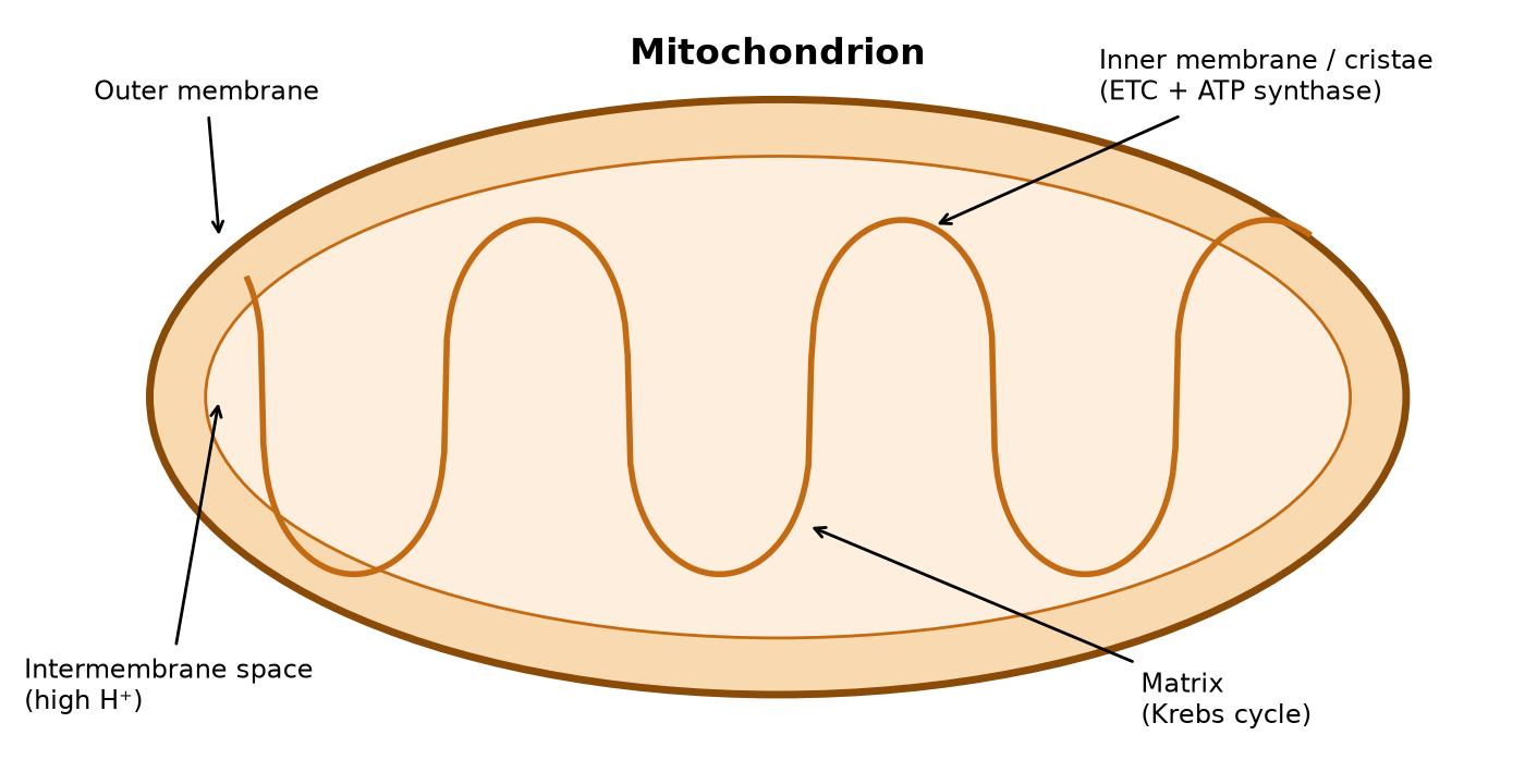
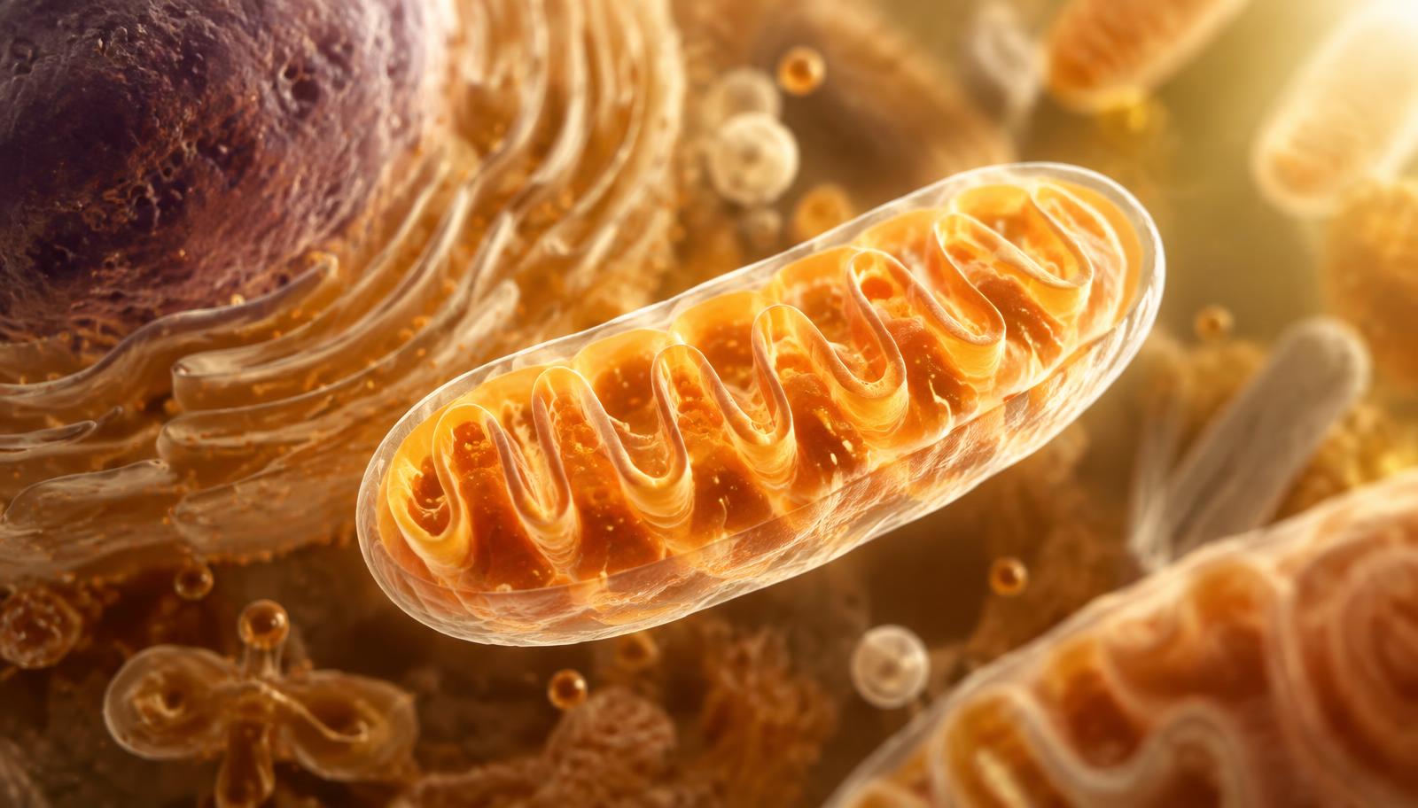
Mitochondria are wrapped in two membranes. The smooth outer membrane forms the boundary, while the inner membrane folds into ridges called cristae. Those folds pack in more surface area for the machinery that makes ATP — the electron transport chain and ATP synthase.
The inner membrane also acts as a wall. It keeps the matrix (where the Krebs cycle runs) apart from the narrow intermembrane space. Because protons (H⁺) can't drift freely across it, the cell can build up a store of them on one side — like water behind a dam.
02 · Energy demand
Busy cells carry more mitochondria
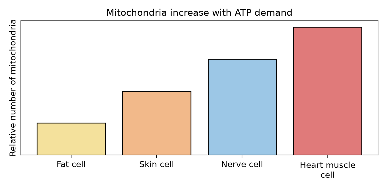
Not every cell needs the same amount of energy. A white fat cell mostly stores fuel, so it gets by with relatively few mitochondria. Skin cells need energy for growth and repair, but not nonstop.
Nerve cells constantly run pumps to keep their electrical signals ready, and heart muscle cells contract every second of your life — so they're packed with mitochondria. The rule of thumb: more ATP demand, more mitochondria. (Brown fat is a fun exception — it's loaded with mitochondria to generate heat.)
03 · Breaking down glucose
From sugar to carbon dioxide
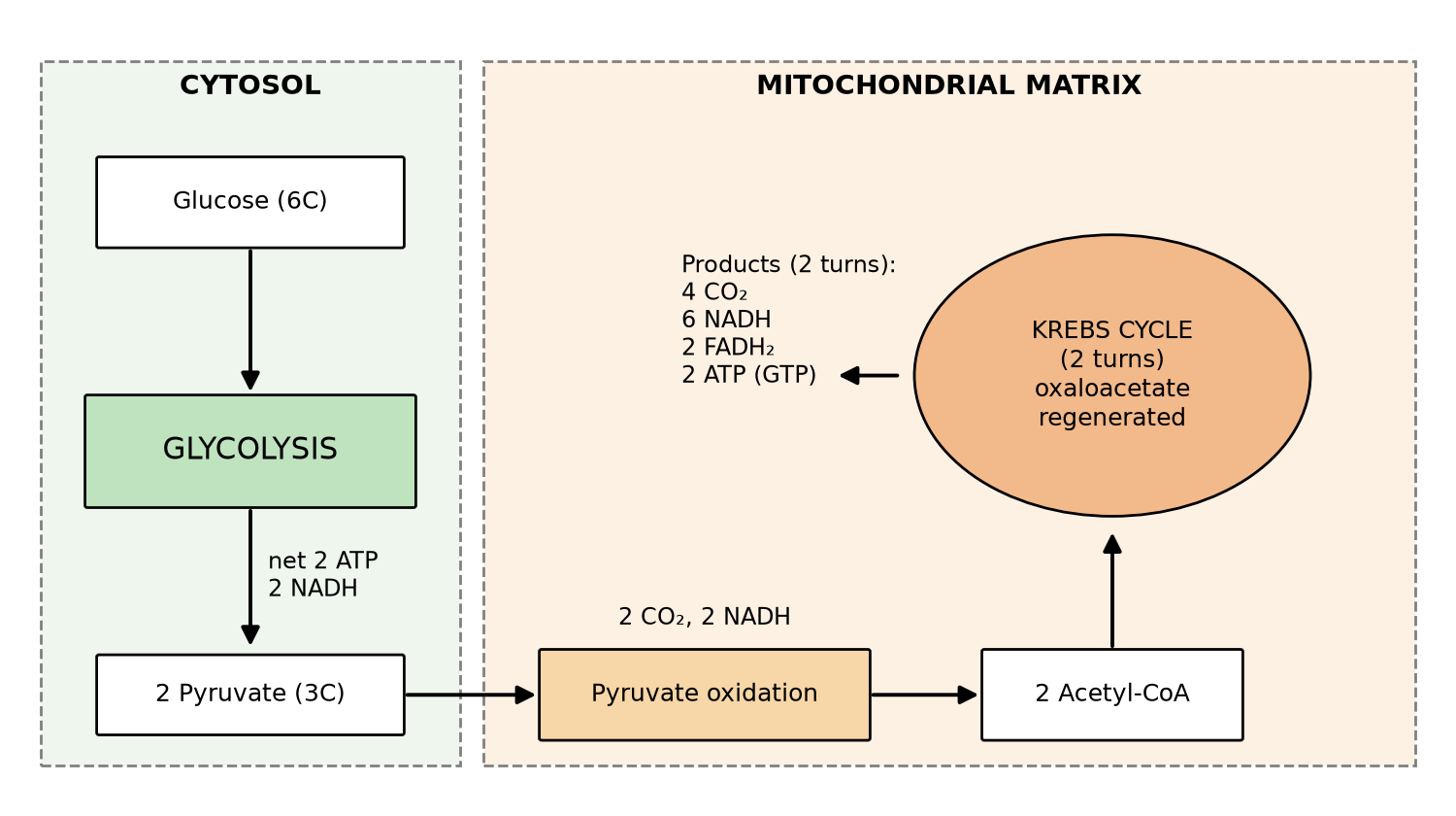
Every glucose molecule starts its journey in the cytosol — the fluid outside the mitochondrion. There, it gets split in half. No oxygen is needed for this step, and no carbon dioxide is released yet. Only a little energy is captured along the way, and most of it is handed to carrier molecules to use later.
The two halves then enter the mitochondrion, get trimmed down, and feed the Krebs cycle — which works like a revolving door: carbon keeps entering, gets stripped away and breathed out as CO₂, and each lap loads more energy onto carriers. By the end, nearly all of the sugar's carbon has left as exhaled CO₂, while most of its energy is still travelling, waiting for the final stage.
04 · The finale
Pumping protons to make ATP
NADH and FADH₂ drop off their high-energy electrons at the start of a chain of proteins embedded in the inner membrane. As the electrons hop from one protein to the next, each one uses that released energy to pump H⁺ into the intermembrane space — building up the "reservoir" from the dam comparison earlier.
Oxygen waits at the end of the chain, accepting the spent electrons and combining with H⁺ to form water — which is why we breathe. With the gradient built, protons rush back through ATP synthase, spinning it like a turbine to stamp out ATP. This flow-driven production is called chemiosmosis, and it's where almost all of the cell's ATP comes from.
05 · The payoff
Adding up the ATP
Where the ATP actually comes from. Tap a segment.
Here's the surprising part: the two early stages bank only a handful of ATP directly. Their real job is to load up the carrier molecules — NADH and FADH₂ — with as much energy as they can carry.
Those carriers then feed the final stage on the inner membrane, where the overwhelming majority of the ATP is actually stamped out. All told, breaking down one glucose molecule yields roughly thirty ATP — plenty to keep a cell running through countless molecular jobs.
The whole story in one line:
sugar + oxygen → carbon dioxide + water + energy (ATP)
Key terms
- ATP
- The cell's energy currency — a molecule that releases energy when it loses a phosphate.
- NADH & FADH₂
- Electron carriers that shuttle energy from glucose to the electron transport chain.
- Cristae
- Folds of the inner membrane that increase surface area.
- Chemiosmosis
- Making ATP using the flow of H⁺ back across a membrane.